
Imaging techniques for retinal diseases
Retinal imaging is an area that plays a critical and increasingly important role in everyday ophthalmic clinical practice, both in diagnosis and therapy management. Interestingly, not only do common eye diseases such as diabetic retinopathy, age-related macular degeneration (AMD) or glaucoma manifest in the back of the eye, but retinal imaging can also be used to draw further conclusions in patients with systemic diseases.
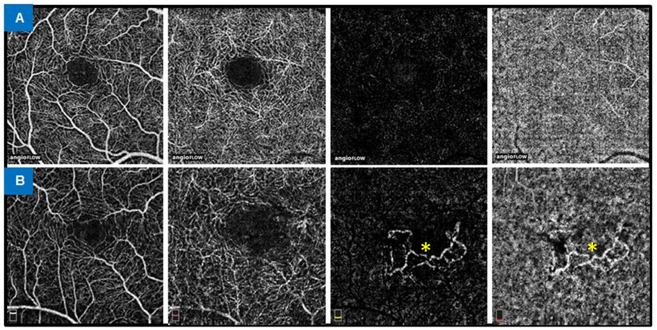
Since the introduction of optical coherence tomography (OCT), imaging techniques have gained tremendous importance in patient care and clinical research. The most recent development in retinal imaging is OCT angiography (OCT-A), which allows non-invasive visualization of retinal vessels at different levels without the use of contrast agents. We are currently pursuing a variety of research interests in this area. On the one hand, we compare the new method with established techniques and analyze the image quality. On the other hand, we are focusing on the clinical application of OCT-A in various ophthalmologic diseases to gain new information on pathomechanisms. The "classical" methods (including OCT, fluorescein angiography, fundus autofluorescence) continue to be used in various studies. Through our Reading Center and the UKM Eyenet, we make our experience and expertise in the field of retinal imaging available to you.
- Analysis of the clinical utility of different imaging modalities in the assessment of retinal disease
- Analysis of technical aspects and evaluation of image quality and reproducibility of different techniques
- Testing and comparison of different devices from different manufacturers with different technical requirements
- Optical coherence tomography (OCT)
- Optical coherence tomography angiography (OCT-A)
- Fluorescein angiography (FLA)
- Fundus autofluorescence (FAF)

