
Experimental Ophthalmology
Our laboratory research focuses on neovascular diseases of the retina, retinopathy of prematurity, and neovascular age-related macular degeneration. We are interested in the pathways that lead to neovascularization as well as potential ways to limit it. The role of the intrinsic immune cell of the retina, the microglia, is also of great interest. We have two experimental mouse models for this purpose. In addition to this topic, we are also investigating the usability of nanoparticles for drug delivery, studying the effects of anti-VEGF drugs on microglia in cell culture and in the living mouse, and culturing retinal pigment epithelial cells on nanofiber meshes.
Neovascular disease in the back of the eye
In our research laboratory, we focus on choroidal neovascularization (CNV), which occurs primarily in advanced age-related macular degeneration (AMD), and retinal neovascularization in retinopathy of prematurity. To study the processes involved in CNV, we induce CNV in mice by laser treatment. This allows us to study the progression of CNV, which involves the migration of immune cells, including an inflammatory response, tissue proliferation, and blood vessel ingrowth. On this basis, we are investigating which therapeutic interventions appear to be particularly suitable for attenuating or stopping these processes and thus reducing the extent of CNV.
Premature babies often have to spend time in an incubator with elevated oxygen levels. This slows down the natural formation of blood vessels in the retina of premature infants, resulting in avascular zones. After the premature infants return to normal breathing air, retinal neovascularization occurs, which can lead to fibrovascular proliferation, retinal detachment and vision loss or even blindness. We simulate these phenomena by placing newborn mice in an oxygen atmosphere for a period of time. Again, we are able to study and quantify the pathological processes. We are using this experimental model to test various agents that inhibit pro-angiogenic molecules or pathways and to determine the extent to which physiological angiogenesis occurs in the avascular zones and neovascularization can be inhibited.
Role of microglia and their inhibition in CNV
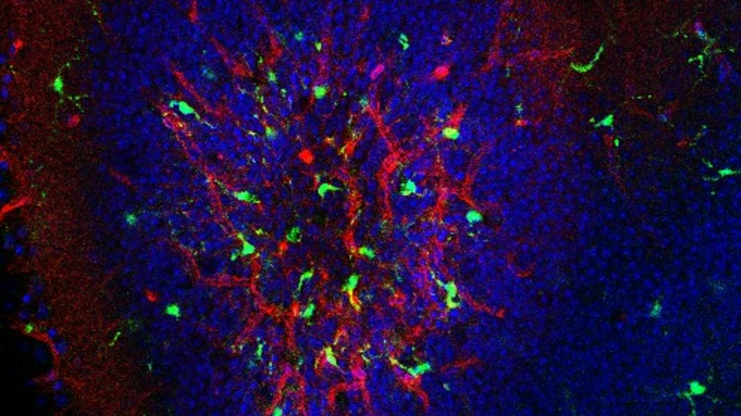
As previously described, immune cells migrate into the area of laser-induced choroidal neovascularization (CNV). This occurs in the acute phase, i.e., up to four days after laser treatment. Most of these immune cells are positive for the marker for phagocytizing monocytes, CD11b. The majority of these cells are microglial cells, the intrinsic immune cells of the central nervous system, including the retina. In addition, a small proportion of migrated peripheral macrophages, dendritic cells and neutrophils are found in the laser foci. These immune cells secrete growth factors and cytokines, such as FGF-2, VEGF, IL-6 and TNF-a, which induce inflammatory responses and neovascularization.
The use of microglial inhibitors such as minocycline significantly reduced the invasion of microglial cells into the laser spot and the leakage at the laser spot, and other substances are being tested. As part of further investigation, we examined the extent to which retinal function is preserved when microglia inhibitors are used. It was found that retinal function was largely preserved when minocycline or the inhibitory tripeptide TKP was used, whereas retinal function deteriorated with the purinergic receptor inhibitor PPADS.
AMD-related growth factors and cytokines
In the model of laser-induced choroidal neovascularization (CNV), a series of complex processes occur in the laser spot after laser treatment, involving a variety of growth factors and cytokines. These are expressed by the various cell populations present in the laser focus, including the aforementioned immune cells, especially microglial cells, but also by the endothelial cells of the neovascular vessels, fibroblasts proliferating in the area of the laser focus, and retinal pigment epithelium (RPE) cells growing back into the laser focus from the edge of the laser focus. We examined the expression of growth factors such as VEGF, PDGF-β, FGF-2, and TGF-β, and cytokines such as IL-1β, IL-6, TNF-β, and CXCL1. We have also examined the expression of some of the receptors for these molecules. On this basis, we are investigating whether and to what extent neovascularization can be inhibited by blocking these growth factors with specific antibodies. While blocking TGF-β has not shown such effects, we have obtained positive results when blocking VEGF, PDGF-β and CXCL1 alone or in combination.
Effects of lipofuscin on microglia and the RPE
One of the functions of retinal pigment epithelial (RPE) cells is to phagocytose and digest the outer segments of photoreceptor cells throughout life. Because RPE cells are post-mitotic, i.e., they no longer divide, non-degradable oxidized and cross-linked lipids and proteins accumulate in them throughout life and collect in former lysosomes. These deposits are called lipofuscin. Outer segment debris not yet taken up by the RPE may also contain components similar to lipofuscin. In addition, RPE cells can release small amounts of lipofuscin as long as the particles are not too large. Since microglial cells migrate into the subretinal space with age, it was of interest to see how they react when confronted with lipofuscin. Therefore, we incubated microglial cells with lipofuscin isolated from human donor eyes and analyzed the resulting behavior of microglial cells in terms of phagocytosis and expression of VEGF and a number of inflammatory cytokines. In particular, the release of IL-6, IL-23p19, RANTES, TNF-a, IL-1a and CXCL1 was stimulated by lipofuscin. Under the influence of minocycline, the release of these cytokines was significantly reduced. Preparations are underway to perform similar studies with RPE cells.
The use of nanoparticles
The effective delivery of drugs remains a particular challenge. One way to deliver drugs in high concentrations to the desired site in the body is to encapsulate them in nanoparticles. The nanoparticles serve as "containers" for the active ingredients and ideally release the active ingredients only at the intended site. In collaboration with researchers at the Agricultural University of Georgia in Tbilisi, we are investigating the properties of nanoparticles in terms of uptake into cultured cells, penetration through ocular tissues and the ability to transport entrapped drugs. The nanoparticles consist of biodegradable pseudoproteins with polyester amides of sebacic acid, leucine and hexanediol as the basic building block. Fluorescein diamide, rhodamine or Nile red are used as dyes and dexamethasone as the active ingredient. The chemical and physical properties of the nanoparticles have already been characterized. Investigations are being carried out in cell cultures (microglia, retinal pigment epithelium) and in the eyes of mice. We have already shown that the nanoparticles penetrate the eye after topical administration. In cell culture, they are taken up by the cells. Further studies will focus on the therapeutic efficacy of nanoparticles loaded with dexamethasone.
Cultivating RPE cells on nanofiber meshes
In a number of eye diseases, the retinal pigment epithelium (RPE), which lies between the retina and the choroid, loses its function and dies. As a direct result, the photoreceptors overlying the RPE also degenerate, leading to visual field defects. For this reason, the question of whether defective RPE can be replaced has been investigated for several years. Autologous transplants or the introduction of a suspension of pigment epithelial cells (retina or iris) into the subretinal space have been attempted. The most promising option seems to be to cultivate the RPE cells to be implanted on a polymeric support, a film, a membrane or a mesh and to place them on this support under the retina. We therefore investigated this approach by culturing RPE cells on nanofiber meshes. These nanofiber meshes, consisting of a copolymer of polycaprolactone and chitosan, were prepared by electrospinning and provided to us by Prof. Dr. Fuchsluger (Rostock). It was shown that RPE cells adhere to these networks and initially upregulate and secrete a number of inflammatory cytokines. The number of adherent RPE cells and the duration of adhesion were significantly improved when collagen was added to the nanofibers. In initial pilot tests, RPE cells adhered at a high density for more than half a year.
Effect of anti-VEGF drugs on retinal microglia
Patients with neovascular AMD have been treated with anti-VEGF agents for several years. For the majority of patients, this is effective in preventing further progression of neovascularization and the edema that forms in the retina during the course of the disease. However, after prolonged use of anti-VEGF medication, some affected eyes experience diminished efficacy and geographic atrophy, or degeneration of the RPE and overlying photoreceptors. To better understand the consequences of anti-VEGF treatment at the cellular level, we isolated and cultured porcine retinal microglial cells and incubated them with different anti-VEGF drugs. While proliferation was little changed, the ability to phagocytose was greatly reduced. Since phagocytosis of degradation products is an important process in the retina, this could be a reason for late effects of anti-VEGF drugs. We are therefore investigating whether microglial cells can be stimulated to regain their phagocytic ability.