Analyse der Blut-Hirn-Schranke von Drosophila

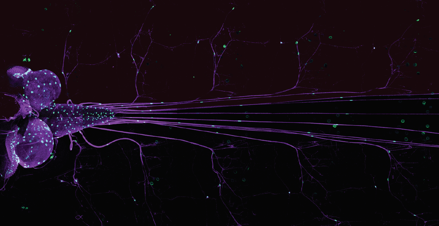
    Die Blut-Hirn-Schranke in Drosophila besteht aus einem Satz spezialisierter Zellen, Septate Junction bildender Zellen. Wir analysieren die Rolle der tricellulären Verbindungen für die Dynamik dieser Zell-Zell Verbindungen während der Bildung und der transienten Öffnung dieser Barriere im Entzündungsfall. Nachdem wir das Einwandern von Makrophagen über die Blut-Hirn-Schranke gezeigt haben, wollen wir jetzt die Signale bestimmen, die die Makrophagen ins Gehirn locken.

    Forschungsgebiet: Neurobiologie

    Prof. Dr. rer. nat. Christian Klämbt  (07/2012 - 06/2024)
    Prof. Dr. rer. nat. Stefan Luschnig   (07/2016 - 06/2024)
    Dr. Stefanie Schirmeier                      (07/2016 - 06/2024; ab 2021 an der Technischen Universität Dresden)
     

    Projektlaufzeit: Juli 2012 - Juni 2024

    • Publikationen

      Originalartikel

      • Schleutker R, Luschnig S (2024) Palmitoylation of proteolipid protein M6 promotes tricellular junction assembly in epithelia of Drosophila. J Cell Sci 137(6): jcs261916. doi:10.1242/jcs.261916
      • Chandran L, Backer W, Schleutker R, Kong D, Beati SAH, Luschnig S, Müller HJ (2023) Src42A is required for E-cadherin dynamics at cell junctions during Drosophila axis elongation. Development. 150(2):dev201119. doi:10.1242/dev.201119
      • Contreras EG, Klämbt C (2023) The Drosophila blood-brain barrier emerges as a model for understanding human brain diseases. Neurobiol Dis. 180:106071. doi:10.1016/j.nbd.2023.106071
      • Glashauser J, Camelo C, Hollmann M, Backer W, Jacobs T, Sanchez JI, Schleutker R, Förster D, Berns N, Riechmann V, Luschnig S (2023) Acute manipulation and real-time visualization of membrane trafficking and exocytosis in Drosophila. Dev Cell. 58(8):709-723.e7. doi:10.1016/j.devcel.2023.03.006
      • McMullen E, Hertenstein H, Müller S, Schirmeier S (2022) Genetically Encoded Sensors to Study Metabolism in Drosophila. Methods Mol Biol. 2540:401-414. doi:10.1007/978-1-0716-2541-5_21
      • Pogodalla N, Winkler B, Klämbt C (2022) Glial Tiling in the Insect Nervous System. Front Cell Neurosci. 16:825695. doi: 10.3389/fncel.2022.825695.
      • Hertenstein H, McMullen E, Weiler A, Volkenhoff A, Becker HM, Schirmeier S (2021) Tarvation-induced regulation of carbohydrate transport at the blood-brain barrier is TGF-β-signaling dependent. Elife. 10:e62503. doi:10.7554/eLife.62503
      • Isasti-Sanchez J, Münz-Zeise F, Lancino M, Luschnig S. (2021) Transient opening of tricellular vertices controls paracellular transport through the follicle epithelium during Drosophila oogenesis. Dev Cell. 56(8):1083-1099.e5. doi:10.1016/j.devcel.2021.03.021
      • McMullen E, Weiler A, Becker HM, Schirmeier S (2021) Plasticity of Carbohydrate Transport at the Blood-Brain Barrier. Front Behav Neurosci. 14:612430. doi:10.3389/fnbeh.2020.612430
      • Winkler B, Funke D, Benmimoun B, Spéder P, Rey S, Logan MA, Klämbt C (2021) Brain inflammation triggers macrophage invasion across the blood-brain barrier in Drosophila during pupal stages. Sci Adv. 7(44):eabh0050. doi:10.1126/sciadv.abh0050
      • Wittek A, Hollmann M, Schleutker R, Luschnig S (2020) The Transmembrane Proteins M6 and Anakonda Cooperate to Initiate Tricellular Junction Assembly in Epithelia of Drosophila. Curr Biol. 30(21):4254-4262.e5. doi:10.1016/j.cub.2020.08.003
      • Sauerwald, J., Backer, W., Matzat, T., Schnorrer, F., and Luschnig, S (2019) Matrix metalloproteinase 1 modulates invasive behavior of tracheal branches during ingression into Drosophila flight muscles. eLife. Elife 2;8. pii: e48857. doi: 10.7554/eLife.48857.
      • Petri, J., Syed, M. H., Rey, S. and Klämbt, C. (2019). Non-Cell-Autonomous Function of the GPI-Anchored Protein Undicht during Septate Junction Assembly. Cell Rep 26, 1641–1653.e4.
      • Dlugos CP, Picciotto C, Lepa C, Krakow M, Stöber A, Eddy ML, Weide T, Jeibmann A, P Krahn M, Van Marck V, Klingauf J, Ricker A, Wedlich-Söldner R, Pavenstädt H, Klämbt C, George B (2019) Nephrin Signaling Results in Integrin beta1 Activation. JASN 30: 1006-1019.
      • Babatz, F., Naffin, E. and Klämbt, C. (2018). The Drosophila Blood-Brain Barrier Adapts to Cell Growth by Unfolding of Pre-existing Septate Junctions. Developmental Cell 47, 697–710.e3.
      • Volkenhoff, A., Hirrlinger, J., Kappel, J. M., Klämbt, C. and Schirmeier, S. (2018). Live imaging using a FRET glucose sensor reveals glucose delivery to all cell types in the Drosophila brain. J Insect Physiol 106, 55–64.
      • Yildirim, K., Petri, J., Kottmeier, R. and Klämbt, C. (2018). Drosophila glia: Few cell types and many conserved functions. Glia 21, 276.
      • Zülbahar, S., Sieglitz, F., Kottmeier, R., Altenhein, B., Rumpf, S. and Klämbt, C. (2018). Differential expression of Öbek controls ploidy in the Drosophila blood-brain barrier. Development 145, dev164111.
      • Misra, T., Baccino-Calace, M., Meyenhofer, F., Rodriguez-Crespo, D., Akarsu, H., Armenta-Calderón, R., Gorr, T.A., Frei, C., Cantera, R., Egger, B. and Luschnig, S. (2017). A genetically encoded biosensor for visualising hypoxia responses in vivo. Biology Open 6, 296–304.
      • Matzat T, Sieglitz F, Kottmeier R, Babatz F, Engelen D, Klämbt C (2015) Axonal wrapping in the Drosophila PNS is controlled by glia-derived neuregulin homolog Vein. Development 142: 1336-1345.
      • Volkenhoff A, Weiler A, Letzel M, Stehling M, Klämbt CSchirmeier S (2015) Glial glycolysis is essential for neuronal survival in Drosophila. Cell Metab 22: 437-447.
      • Limmer S, Weiler A, Volkenhoff A, Babatz F, Klämbt C (2014) The Drosophila blood-brain barrier: development and function of a glial endothelium. Front Neurosci 8: 365. [Stefanie Schirmeier, neé Limmer]

      Reviews

      • Schirmeier S, Matzat T, Klämbt C (2016) Axon ensheathment and metabolic supply by glial cells in Drosophila. Brain Res 1641(Pt A): 122-129. (doi: 10.1016/j.brainres.2015.09.003. Epub 2015 Sep 12.)